Posterior Fossa Vascular Supply From Eye of Rhoton: Recapitulation (Part1)
Three arteries of Posterior Fossa namely Superior Cerebellar Artery(SCA), Anterior Inferior Cerebellar Artery (AICA) and Posterior Inferior Cerebellar Artery (PICA) bear a consistent relationship to other structures occurring in 3 neurovascular complexes.
An upper complex related to the SCA, a middle complex related to the AICA, and a lower complex related to the PICA.
Each neurovascular complex includes one of the 3 parts of the brainstem, one of the 3 surfaces of the cerebellum, one of the 3 cerebellar peduncles, and one of the 3 major fissures between the cerebellum and the brainstem.
In addition, each neurovascular complex contains a group of CNs. The upper complex includes the oculomotor, trochlear, and trigeminal nerves that are related to the SCA.
The middle complex includes the abducent, facial, and vestibulocochlear nerves that are related to the AICA.
The lower complex includes the glossopharyngeal, vagus, accessory, and hypoglossal nerves that are related to the PICA.
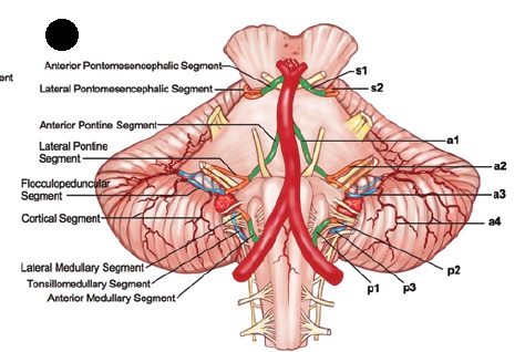
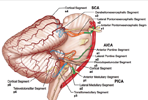
According to Rhoton’s proposed scheme cerebellar arteries were designated by the first letter of the artery’s name: s denotes the SCA; a, the AICA; and p, the PICA.
Lowercase letters were used to distinguish cerebellar arteries from cerebral arteries with the same first letter of the artery’s name (specifically, ACA and PCA). The segmental anatomy was numbered in ascending order from proximal to distal segments.
Super Cerebellar Artery
- The SCA arises in front of the midbrain, usually from the basilar artery near the apex, and passes below the oculomotor nerve, but may infrequently arise from the proximal PCA (Posterioir Cerebral Artery) and pass above the oculomotor nerve.
- The SCA usually arises as a single trunk, but may also arise as a double (or duplicate) trunk.
- The SCAs arising as a single trunk bifurcate into a rostral and a caudal trunk. The rostral trunk supplies the vermian and paravermian area, and the caudal trunk supplies the hemisphere on the suboccipital surface.
- The SCA frequently has points of contact with the oculomotor, trochlear, and trigeminal nerves.
The SCA is divided into 4 segments:
- anterior pontomesencephalic s1
- lateral pontomesencephalic s2
- cerebellomesencephalic s3
- cortical s4
Each segment may be composed of one or more trunks, depending on the level of bifurcation of the main trunk.
Anterior Pontomesencephalic Artery s1
- This segment is located between the dorsum sellae and the upper brainstem.
- It begins at the origin of the SCA and extends below the oculomotor nerve to the anterolateral margin of the brainstem.
- Its lateral part is medial to the anterior half of the free tentorial edge.
(Green Part in diagram)
Lateral Pontomesencephalic Segment, s2
- This segment begins at the anterolateral margin of the brainstem and frequently dips caudally onto the lateral side of the upper pons.
- Its caudal loop projects toward and may reach the root entry zone of the trigeminal nerve at the midpontine level.
- The trochlear nerve passes above the midportion of this segment.
- The anterior part of this segment is often visible above the tentorial edge, but the caudal loop usually carries it below the tentorium.
- This segment terminates at the anterior margin of the cerebellomesencephalic fissure.
(Orange Coloured part in diagram)
Cerebellomesencephalic Segment, s3
- This segment courses within the cerebellomesencephalic fissure.
- The SCA branches enter the shallowest part of the fissure located above the trigeminal root entry zone and again course medial to the tentorial edge with its branches intertwined with the trochlear nerve.
- The fissure in which the SCA proceeds progressively deepens medially and is deepest in the midline behind the superior medullary velum.
- Through a series of hairpin-like curves, the SCA loops deeply into the fissure and passes upward to reach the anterior edge of the tentorial surface, the surface facing the lower margin of the tentorium. The trunks and branches of the SCA are held in the fissure by branches that penetrate the fissure’s opposing walls.
(Blue Coloured part in diagram)
Cortical Segment, s4
This segment includes the branches distal to the cerebellomesencephalic fissure that pass below the tentorium and are distributed to the tentorial surface.
(Maroon coloured vessels on tentorial cerebellar surface)
Reference:
https://www.ncbi.nlm.nih.gov/pubmed/21548748


